EYE DISEASES
EYE DISEASES
Being aware of eye diseases is important because early detection and treatment can help prevent vision loss and improve the overall health of the eye.A large percentage of the human population will experience eye problems at some point in their lives.
These are our top 4 eye conditions.
-
![]()
-
![]()
-
![]()
Glaucoma
Glaucoma -
![]()
Retinal Care
Retinal Care


Diabetes
Diabetic Eye Exam
Why it is important to get an eye exam yearly if you are a diabetic:
- Early detection and prevention of ocular diseases, such as diabetic retinopathy.
High blood sugar can cause damage to the blood vessels in your eyes, leading to:
- Blurry vision
- Cataracts
- Glaucoma
- Blindness
- Retinal Hemorrhaging
- Diabetic Retinopathy
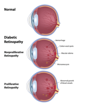
Diabetic Retinopathy
Diabetic retinopathy gets worse over time when left untreated or when blood sugar is not maintained and stable. This will cause the blood vessels on the retina’s surface to break and pull away from their normal position inside the eye. This will then lead to a retinal detachment, which requires immediate medical attention.
The Two Types of Diabetic Retinopathy:
Proliferative Diabetic Retinopathy: When the condition becomes more advanced, patients will often begin to notice more visual disturbances. This happens because the blood vessels around the retina start to grow abnormally.
Non-Proliferative Diabetic Retinopathy: This is the earliest stage of diabetic retinopathy, patients may experience mild to no symptoms. This happens because the blood vessels have started to become weak, but have not broken yet.
Symptoms and Risk Factors
It is important to make sure that you are maintaining and treating your diabetes properly to prevent ocular diseases or the progression of them. Our specialist recommends getting a yearly eye exam to accurately diagnose and treat symptoms and diseases related to diabetes. Symptoms that indicate damage related to diabetes include:
- Visual Disturbances, such as blurriness
- Halos, floaters, or shadows that were not there prior to the diagnosis of diabetes
- Ocular pain
- Early development of cataracts
- Neovascular glaucoma - which is a rare type of glaucoma that develops when blood vessels grow abnormally on the iris (colored part of the eye).
- Retinal hemorrhaging
People that have Type 1, Type 2, and gestational diabetes are all at risk for developing ocular diseases, such as diabetic retinopathy, and should get their eyes examined regularly to help prevent these diseases.
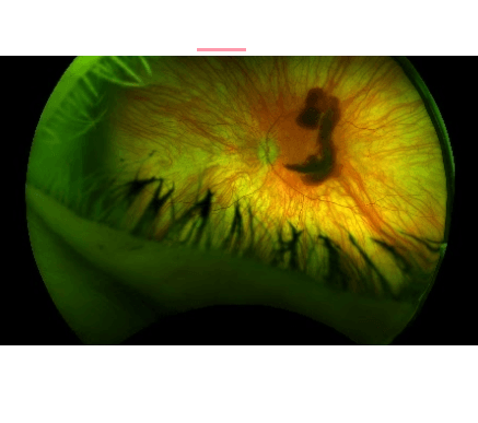
The image shows a retinal hemorrhage in a patient that has fluctuating blood sugar levels and has not properly maintained their diabetes.
Caring for our Diabetic Patients
Dr. Nicole Kish is our ocular disease specialist. She helps to diagnose, treat, and prevent ocular diseases like diabetes in the eye. She, along with trained technicians in the office will use advanced technology and diagnostic tools to examine your eyes for any symptoms that might indicate further testing or treatment. Dr. Kish will then be able to clarify any information regarding your symptoms and treatment options. We care about our patients and it is important to us that you receive the best care possible.
If you currently do not have a primary care physician, Dr. Nicole Kish can help to recommend you to a specialist to help with the management or treatment of your diabetes.
Cataracts
What are cataracts?
Having cataracts can make it seem like you’re seeing the world through a dusty windshield. The clouding of your eye’s natural lens, which is behind the iris and pupil, is called a cataract. Cataracts typically develop with age and are one of the most common eye conditions we treat at Dr. Nicole Kish’s office. Protecting your eyes from the sun and not smoking can help slow down the development of cataracts, but most people will eventually get them.
Treating Cataracts
The lens of the eye helps focus light on the retina, the tissue at the back of the eye. When the lens has become thickened and cloudy, the only effective treatment is surgically removing the cataract. Cataract removal is a common procedure and it’s safely performed millions of times each year.
Symptoms of Cataracts
Aging is the most common cause of cataracts, which usually develop gradually over several years. Cataracts in younger people or diabetics tend to develop more quickly. When cataracts develop, the cloudiness decreases your visual acuity and produces a variety of symptoms, including:
- Blurry vision
- Seeing double
- Light sensitivity
- Problems seeing at night
- Bright colors appear dull
- Halos surrounding lights
- Increased sensitivity to glare
How to slow down your development of cataracts:
Protecting your eyes is the best way to do this. Wear sunglasses that screen out the sun’s ultraviolet (UV) light rays. You may also wear regular eyeglasses that have a clear, anti-UV coating. Talk with your eye doctor to learn more.
Subcapsular cataract: This type occurs at the rear of the lens. It’s most prevalent in people with diabetes or people who take high doses of steroids medications.
Different Types of Cataracts
There are 3 main types of cataracts
Nuclear cataract: This type develops deep within the nucleus of the lens. Nuclear cataracts are the most common age-related cataracts.
Cortical cataract: Characterized by whitish cloudy areas, these cataracts develop on the outside edge of the lens, called the cortex.
Other cataracts:
- Congenital cataracts, which are present at birth or form during a baby’s first year, are less common than age-related cataracts.
- Secondary cataracts are caused by disease or medications. Diseases that are linked with the development of cataracts include glaucoma and diabetes. The use of the steroid prednisone and other medications can sometimes lead to cataracts.
- Traumatic cataracts develop after an injury to the eye, but it can take several years for this to happen.
- Radiation cataracts can form after a person undergoes radiation treatment for cancer.
Surgery
Cataract surgery can be performed using a small blade (traditional cataract surgery) or with the assistance of a laser. Traditional cataract surgery is one of the most common surgical procedures performed in the world and is often the most appropriate choice for patients. Cloudy, blurry vision often indicates the presence of cataracts, especially in men and women over age 50. Our practice includes one of the best cataract surgeons, Dr. Brian Morgan.
Traditional Cataract Surgery
During traditional cataract surgery, your ophthalmologist makes a tiny incision and then inserts a pen-shaped probe that uses ultrasound energy to break up the cloudy lens into tiny pieces. This process is called phacoemulsification. The probe then suctions the pieces from the eye. The eye surgeon then replaces the cloudy lens with an artificial intraocular lens (IOL). Different types of IOLs are available and our ophthalmologists clearly explain the differences prior to your surgery.
Laser Cataract Surgery
Laser cataract surgery uses laser energy to create an incision in the cornea, but most of the procedure is quite similar to traditional cataract surgery. Not all patients are appropriate candidates for laser cataract surgery. Your ophthalmologist reviews the options during your initial appointment and gathers your medical history and specific vision concerns before recommending the treatment plan best suited for you.
Cataract surgery is an outpatient procedure that typically takes about 10 minutes, whether it’s traditional or laser-assisted surgery.
YAG Laser Procedure
Problems after cataract surgery are rare but can occur. Sometimes the tissue that encloses the artificial intraocular lens becomes cloudy and blurs the vision. This is known as a secondary cataract, even though it’s not a true cataract. This can develop months or even years after cataract surgery. This outpatient procedure is treated with a laser called YAG laser capsulotomy
How long does it take to recover from cataract Surgery?
The recovery period for both laser-assisted cataract surgery and traditional cataract surgery is the same. Some people can see clearly almost immediately, while others may find their vision clears within about a week or two. Remember that it takes about 3 months to fully recover from cataract surgery.
Glaucoma
What is glaucoma?
Glaucoma is actually a group of eye diseases that damage the optic nerve and result in the loss of vision if left untreated. Even though elevated eye pressure is one of the main risk factors for glaucoma, up to one-third of men and women diagnosed with glaucoma don’t have a history of high eye pressure. Glaucoma occurs when the eye is vulnerable to nerve damage. Normally, the anterior chamber of the eye is filled with fluid that continually flows in and out through the open angle where the cornea and iris meet. Your eyes typically regulate the amount of pressure inside through this drainage.
Glaucoma often occurs when this pressure is higher than normal, but it can also happen when the eye pressure is considered normal. When glaucoma isn’t diagnosed it can cause serious damage to the optic nerve and result in loss of vision and even blindness
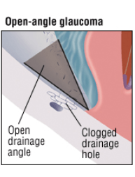
Open-Angled Glaucoma
This is a common type of glaucoma, 90% of Glaucoma cases are open-angle. It is one that gradually erodes your peripheral vision causing blind spots and “tunnel vision”. Open-angle glaucoma involves a gradual increase of eye pressure that develops for years due to clogged canals, our office persistently checks patients' pressure with the tonometry machine with every visit to observe any changes with patients' pressure. Open-angle glaucoma is caused by the slow clogging of drainage canals with wide or open angles between the iris and cornea and causes swelling of the optic nerve. Permanent damage can occur before it’s diagnosed because it develops slowly, that’s why glaucoma is sometimes called the “silent thief of sight.” Undergoing a comprehensive eye exam at least once every year can help detect many types of glaucoma during its early stages. For patients with a family history of glaucoma, suspect of glaucoma, or has glaucoma it is very crucial to come in for regular visits to monitor any changes.
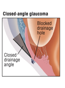
Closed-Angled Glaucoma
A sudden and sharp increase in intraocular pressure-commonly referred to as a glaucoma attack is a medical emergency that requires immediate treatment. This happens when the drainage system of the eye is completely blocked and fluid cannot be drained from the eye. In addition to sudden loss of vision, narrow-angle glaucoma can cause headaches, glare and halos, red eyes, nausea, and vomiting. Untreated eyes have the potential to lead to blindness, require immediate medical attention, and possibly surgery.
Treatment for Glaucoma
Most people diagnosed with glaucoma respond well to eye drops with few side effects. Glaucoma eye drops used to cause blurred vision, but this is no longer the case. The doctor will recommend eye drops based on the patient’s medical history, and in some cases, more than one eye drop may be prescribed. Depending on the patient's preference and the severity of the disease, our doctor may recommend one of the following alternative treatments:
- Glaucoma Eye Drops to lower pressure in the eyes and maintain stability.
- Laser eye surgery to drain fluid from the eyes. It may be performed in addition to taking glaucoma medicine.
- Conventional eye surgery creates a new channel for fluid to drain from the eye. This option is typically recommended only if patients don’t respond to medication and laser eye surgery.
Retinal Care
About the Retina
Every image we see passes through the lens of the eye and is absorbed by the retina. The retina converts the image into an electrical signal and sends it to the brain. A healthy retina is essential for vision.
The eye works much like the lens of a camera. Light is reflected off an object then passes through the cornea and lens of your eye. The cornea and lens are responsible for bending this light so that it lands directly onto the retina. The retina is the nerve layer that senses light and sends impulses through the optic nerve to the brain about what has been seen.
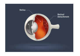
Retinal Detachment
Retinal detachment is a medical emergency that requires immediate treatment. A torn or detached retina is often the result of injury, but it can also occur spontaneously, especially among older men and women. A detached retina is separated from the structures that hold it in place and surgery is needed to reattach it. Symptoms of a detached retina that’s not caused by trauma include blurry vision or floaters, which may steadily increase.
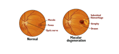
Age-Related Macular Degeneration (AMD)
Age-related macular degeneration is the main cause of vision loss. It occurs when the central portion of the retina, called the macula, deteriorates. This causes central vision loss. You may have heard of “dry” and “wet” forms of AMD. Dry AMD is much more common and treatable compared to the much rarer wet form of AMD, which can cause rapid vision loss. Common macular degeneration symptoms include a blind spot in your central vision or distortion of the central vision. Routine eye exams can detect the condition even before you notice any symptoms.
Diabetic Retinopathy
We provide eye care for a range of diabetes-related eye diseases. These conditions are a risk of people with type 1, type 2, or gestational diabetes. Diabetic retinopathy occurs when the blood vessels in the eye weaken, break down, and form scar tissue on the retina. The best defense against developing this condition is prevention by managing your diabetes. Our optometrist can help you understand the risk of diabetic retinopathy and other eye diseases.